Blood Cancer & COVID-19
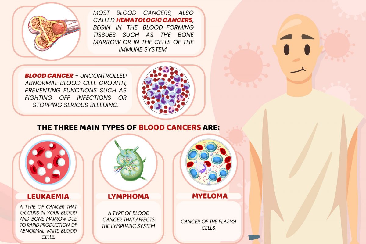
Most blood cancers, also called hematologic cancers, begin in the blood-forming tissues such as the bone marrow or in the cells of the immune system.1 Blood cancers occur when the normal blood cell development process is interrupted by uncontrolled abnormal blood cell growth, preventing functions such as fighting off infections or stopping serious bleeding.2
The three main types of blood cancers are2:
- Leukemia – a type of cancer that occurs in your blood and bone marrow due to rapid production of abnormal white blood cells. The high level of abnormal white blood cells are not able to fight off infections and cause impairment in the production of red blood cells and platelets in the bone marrow.
- Lymphoma – a type of blood cancer that affects the lymphatic system; which removes excess bodily fluids and produces immune cells. Lymphocytes are white blood cells that fight off infection. Abnormal lymphocytes turn into lymphoma cells that multiply and collect in your lymph nodes and other tissue which overtime, will impair your immune system.
- Myeloma – cancer of the plasma cells. Plasma cells are white blood cells that produce antibodies to fight off diseases and infections in your body. Myeloma cells prevent the normal production of antibodies causing the body’s immune system to be weakened and susceptible to infection.
Blood cancer & COVID-19
As COVID-19 emerged, patients with blood cancers are at increased risk of severe COVID-19 infection and high mortality rate due to their compromised immune system and inability to receive necessary care in time.3, 4, 5, 6 Medical practitioners have to assess whether treatment plans should begin on schedule or delayed until a certain time.5, 6 Studies have shown that patients with blood cancers are more susceptible to severe COVID-19 infection than patients with solid cancers or healthy people, likely due to the immunosuppressive nature of their disease and/or treatments. However, researchers sought to discover if immunosuppression caused patients with blood malignancies to have a decreased immune response to the COVID-19 vaccines that are now being provided around the world.6
To refer for Pantai Premier Pathology tests related to Blood Cancer, please click on the below button.
CLICK HERE
For more information on the tests provided, please contact us at +603-42809115 (Customer Service) or email us at info@premierpathology.com.my
References
1- hematologic cancer. (n.d.). National Cancer Institute (NIH). Retrieved May 4, 2021, from https://www.cancer.gov/publications/dictionaries/cancer-terms/def/hematologic-cancer
2- Blood Cancers. (n.d.). American Society of Hematology. Retrieved May 4, 2021, from https://www.hematology.org/education/patients/blood-cancers
3- Ferrara, F., Zappasodi, P., Roncoroni, E., Borlenghi, E., & Rossi, G. (2020). Impact of Covid-19 on the treatment of acute myeloid leukemia. Leukemia, 34(8), 2254-2256.
4- He, W., Chen, L., Chen, L., Yuan, G., Fang, Y., Chen, W., … & Gale, R. P. (2020). COVID-19 in persons with haematological cancers. Leukemia, 34(6), 1637-1645.
5- Burki, T. K. (2020). Cancer guidelines during the COVID-19 pandemic. The Lancet Oncology, 21(5), 629-630.
6- American Association for Cancer Research. (2021, February). Survival Factors ID’d for Patients with Blood Cancer + COVID-19. Cancer Discovery. https://cancerdiscovery.aacrjournals.org/content/11/2/214.2