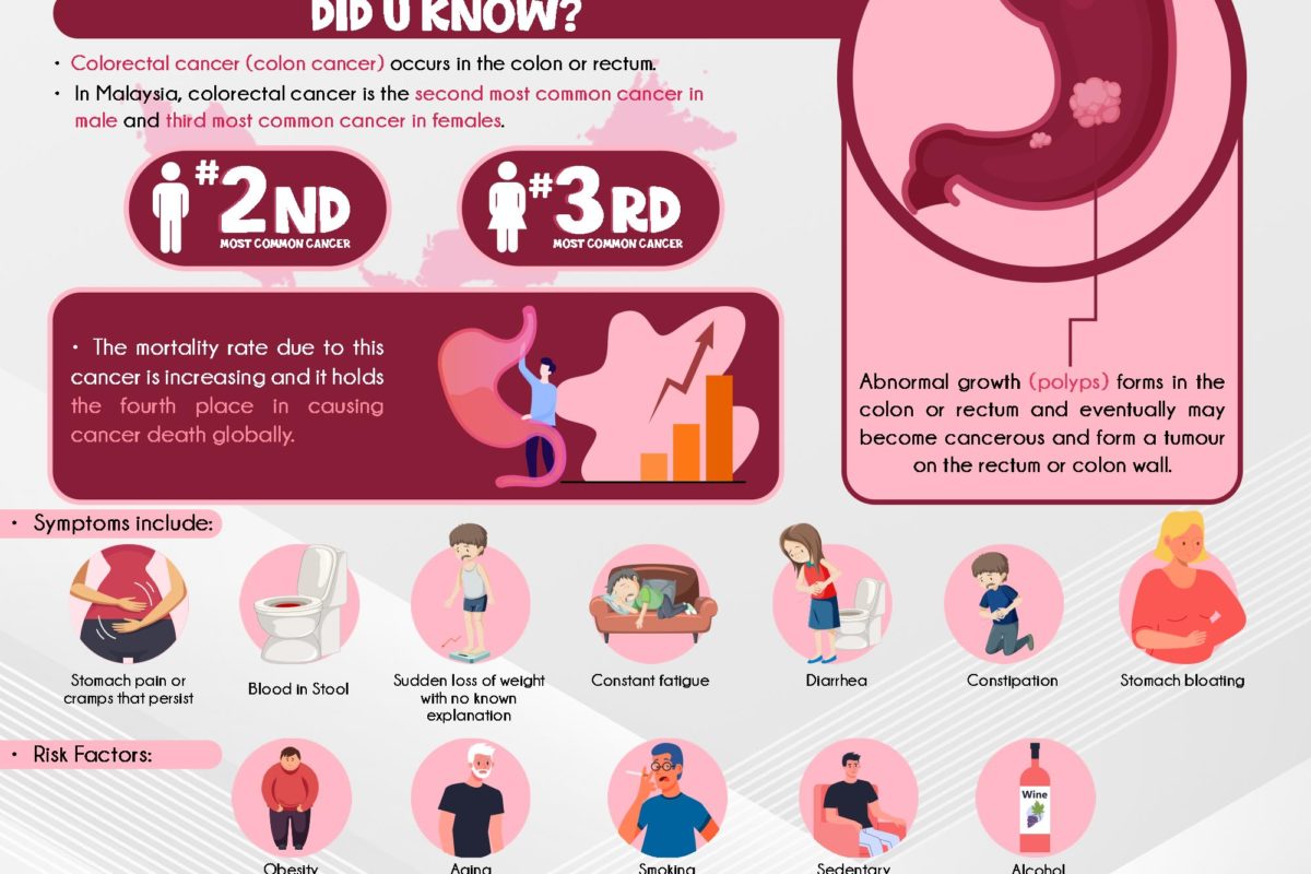
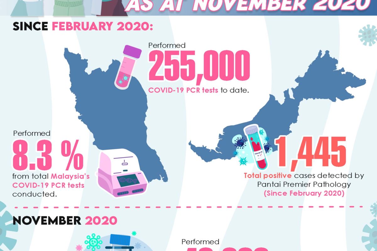
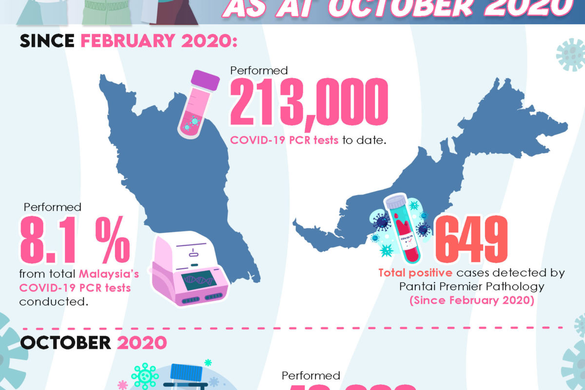
Health screening is an efficient way to detect diseases early, especially when there are no signs or symptoms of the diseases present. It is an important routine medical examination performed to ensure optimal health for everyone as it allows patients to have better control over their health. Detecting a disease or condition early helps to prevent the development of chronic disorders and avoid complications.
Haematology
Haematology laboratory testing is performed as part of a blood test package for health screening. It involves testing the blood and its constituents to determine how blood can affect overall health or disease. The tests can also be used to diagnose numerous conditions such as inflammation, anaemia, infection, allergies, haemophilia, blood-clotting disorders, leukaemia and the body’s response to chemotherapy. The blood test results can provide an accurate analysis of the patient’s body condition and the role of both the internal and external influences that may affect a patient’s health.1
Blood banking refers to the collection, separation and storage of blood so that they can be used according to patients’ needs. ABO grouping and RhD grouping is done to ensure that a recipient receives compatible blood during transfusion.2
Antigens within the ABO groups are what determines an individual’s blood type. The four main blood groups are blood group A – presence of A antigens on red blood cells with anti-B antibodies in plasma; blood group B – presence of B antigens with anti-A antibodies in plasma; blood group O – no antigens present with both anti-A and anti-B antibodies in plasma; and blood group AB – presence of both A and B antigens with no antibodies. Receiving blood from the ABO group that does not match can be life threatening.3
The Rh blood grouping is very crucial as Rh antigens are highly immunogenic. An individual who does not produce the D antigen will present the anti-D antibody when they encounter the D antigen during RBC transfusion causing a hemolytic transfusion reaction or hemolytic disease of the new born on fetal RBCs. Therefore, it is important for the Rh status to be determined routinely in blood donors, transfusion recipients and expecting mothers.4
Renal Function Test
The kidneys play important roles in maintaining health by filtering waste materials from the blood and expel them from the body through urine. The kidneys also help in maintaining water and various essential mineral levels in the body.5 Renal function test examines whether the kidneys are healthy and functioning properly by measuring albumin to creatinine ratio (ACR) and glomerular filtration rate (GFR). ACR is a urine test that measures the albumin level in your urine. High levels of albumin in your urine is an early sign of kidney damage, whereas, GFR is a blood test used to follow up patients with known kidney disease by measuring the kidney function to determine the stage of kidney disease (there are 5 stages).6
Gout
Uric acid is a normal by-product that is being made when the body breaks down chemicals known as purine. Purines are found in our own cells and also in some foods. Most of the uric acid dissolves in the blood and then travels to the kidney. It then leaves the body through urine. When the body produces uric acid in excess or not eliminating it enough into the urine, it deposits in the joints and forms crystals. This condition is known as gout which is a form of arthritis that results in painful inflammation around the joints. 7, 8
Uric acid blood test is used to diagnose gout when a patient shows symptoms such as pain and/or swelling in the joints (i.e. big toe, ankle or knee), appearance of red skin around the joints or joints that feel warm upon touch due to inflammation.7
The test is also used to monitor patients with gout who are at a high risk of developing kidney stones.7, 8
Diabetic Screen
The body’s main source of energy comes from blood glucose that is obtained through diet. The glucose is transported to the brain, cells and tissues to be used as energy by insulin, a hormone made by the pancreas. Sometimes the body does not produce adequate or any insulin or is not able to use it. This is when glucose remains in the blood and does not reach the cells.9, 10
Diabetes is a disease that develops when the blood glucose level is too high. Untreated diabetes can damage the eyes, nerves, kidneys and other organs.9
Diabetic screening is used to diagnose diabetes or pre-diabetes. This allows health care professionals to detect diabetes sooner and work with the patients to manage or prevent complications as well as delay or prevent type 2 diabetes.9, 10, 11
HbA1c test is used to monitor the glucose level of patients that have already been diagnosed with diabetes and this may be done every 2-6 months. This test measures the average glucose level which is attached to part of the red blood cells over the past 2-3 months.9, 10, 11
Liver Function Test
The liver carries out many important metabolic functions such as converting nutrients in our diets into biochemicals that can be used by the body, store these biochemicals and provide them to the cells when needed. It also converts toxic substances from the blood and converts them into harmless substances or release them out of the body.12
Liver Function Test is used to:
- Evaluate the biochemicals found in the blood. A higher or lower than normal levels of these enzymes or proteins in the blood implies a problem in the liver. An abnormal result usually requires a follow up to identify the source of the abnormalities
- Diagnose liver disorder if symptoms related to liver disease are observed (i.e. jaundice, abdominal pain and swelling, edema in legs and ankle, etc.)
- Monitor the severity of a previously diagnosed liver disorder
- Test for any potential liver disease (i.e. alcohol-dependent individuals or individuals who have been exposed to hepatitis virus)
- Monitor side effects of some medicines 13
Lipid Profile
Lipids are a type of fat and fat-like substances. They are important elements which make up the cells and are a source of energy. Cholesterol and triglyceride are two important lipids found in the blood.14
While the body produces cholesterol to function properly, diet is found to be the source of some cholesterols which contributes to high blood cholesterol level.15
In the long term, undiagnosed high blood cholesterol level will result in the formation of plaque which blocks the opening of blood vessels that can restrict blood flow (atherosclerosis) in the arteries. This incident has been associated with heart disease and other health conditions.16, 17
Lipid profiling is done to:
- Examine the risk of developing cardiovascular disease
- Alert medical practitioners whether a patient requires any treatment or lifestyle changes to lower blood cholesterol level
- Reduce the patient’s risk ratio to heart disease by monitoring the treatment of unhealthy lipid levels 17
It is important to monitor and maintain these lipids in normal levels to stay healthy.
Urine Examination
(Urine Full Examination & Microscopy Examination- FEME)
The kidneys and bladder work together to remove waste material, minerals and other substances from the blood through urine. Many factors such as diet, fluid intake, kidney function and exercise affect the constituents of urine.18, 19
Urine tests provide information to many underlying diseases and function as a patient’s health indicator. A routine urine test helps:
- To identify the cause for numerous different symptoms of a urinary tract infection which includes:
- Pain when urinating
- Flank pain
- Foul-smelling urine
- Blood in urine (Haematuria)
- Fever
- To monitor the treatment of conditions such as diabetes, kidney stones or urinary tract infection (UTI)
- As part of a routine check-up20
A urine full examination observes abnormalities in the colour, clarity, odour, specific gravity, pH, protein, glucose, ketones, bilirubin, urobilinogen, nitrites, leukocyte esterase. Whereas, a urine microscopic examination is performed:
- To verify the results from the urine analyser when unusual findings with the physical or chemical test are observed
- To detect the amount and or presence of red blood cells, white blood cells, cast, crystals, bacteria or yeast in urine20, 21
Tumour Markers:
Alpha- Fetoprotein (AFP), CA 19.9, Carcino Embryonic Antigen (CEA), CA12.5, CA15.3
Tumour markers are biomarkers which are often proteins found in blood, urine or body tissues. They are produced by cancerous tissue or by the body itself in response to the growth of cancer or certain benign (non-cancerous) conditions. These biomarkers may be used together with other tests (scans, biopsies, imaging, etc) to:
- Determine prognosis- tumour marker level reflects how aggressive (staging) a cancer is likely to be
- Help identify and diagnose several types of cancer at an early stage- an increased level of the circulating tumour markers may indicate the presence of cancer
- Detect and monitor a patient’s response to treatments- a decreased circulating tumour marker level indicates that the patient is responding to the cancer treatment, whereas, an increased or unaltered tumour marker level suggest that the patient is not responding to the cancer treatment
- Detect recurrence- the tumour marker’s level is measured using multiple samples taken at various times during and after treatment (“serial measurements”) which reflects the marker’s level over time
- Obtain details on how aggressive a cancer is (staging) or whether it can be treated with a targeted therapy
- Screen individuals at high risk of cancer- risk factors include family history and specific risk factors of a particular cancer22, 23, 24, 25
The type of test you will receive is based on your health history and symptoms that you may have.
Others: C- Reactive Protein (CRP)
C- reactive proteins (CRP) are a type of proteins made by the liver. The level of CRP increases in the blood when a condition which causes inflammation occurs in the body. CRP is one of the most sensitive and acute phase reactants which means that it is released into the bloodstream within a few hours upon an injury, beginning of an infection or other triggers of an inflammation. A significant increase in the CRP level is observed with:
- Trauma or heart attack
- Fungal infection
- Current or untreated autoimmune disease (i.e. rheumatoid arthritis or lupus)
- Severe bacterial infection (i.e. sepsis)
- Bone infection (i.e. osteomyelitis)
- Inflammatory bowel disease26, 27, 28, 29, 30
The CRP test provides details whether an inflammation is present to the medical practitioner. The test measures the level of CRP in the blood to identify whether the inflammation is an acute condition or to monitor the severity of disorder of a chronic condition which will then be followed-up with additional testing and treatment. If the CRP levels reduce, it’s a sign that the treatment for inflammation is effective. The level of CRP is used as inflammation markers.26, 27, 28, 29, 30
Immunology & Serology
An immunology test will determine if an individual has an autoimmune disease which affects the tissues or organs throughout the body, whereas, a serology test is a blood test which measures the antibodies in the blood. Several types of serological tests are used to diagnose various disease conditions.31
Immunology and serology tests are used to:
- Identify antibodies- antibodies are proteins made by white blood cells in response to a foreign particle (antigen) in the body
- Examine problems with the immune system- i.e. immune system attacks its own tissues (autoimmune disorder) or when the immune system functions improperly (immunodeficiency disease)
- Determine compatibility of organ, tissue and fluid transplantation31
Endocrinology: TSH, Free T4, Free T3, IGF- 1, Cortisol (AM/PM), DHEA- Sulphate, Estradiol, Progesterone, SHBG, Testosterone, Insulin, LH, FSH, Prolactin
The endocrine system is an integrated structure that includes numerous glands found throughout the body. This system together with the nervous system, monitors and regulates various vital internal functions of the body such as influencing heart beats, bone and tissue growth, metabolism, movement, sensory perception and ability to reproduce. The system consists of a network of glands which produces, stores and secretes hormones (chemical signals) into the bloodstream to regulate numerous bodily functions.32, 33
The endocrine feedback system assists in the control of hormonal balance in the bloodstream. When the body has too much or too little of a specific hormone, the feedback system alarms the appropriate gland to fix the problem.33 When the feedback system has trouble to maintain the hormones at the right level or if the body has trouble clearing them from bloodstream, hormonal imbalance occurs.32 Hormonal imbalance may occur due to:
- Tumour or injury of an endocrine gland
- Genetic condition (i.e. multiple endocrine neoplasia (MEN) or congenital hypothyroidism)
- Problem in the endocrine feedback system
- Inability of a gland to stimulate another gland to release hormones
- Infection or severe allergic reactions34
An endocrinology test is used to:
- Measure the various levels of hormones in the patient
- Monitor that the endocrine glands are functioning properly
- Determine the cause of an endocrinological disease
- Confirm an early diagnosis
- Determine if medication or treatment plan needs to be modified34
Additional Test:
Vitamin D
Vitamin D is a fat-soluble vitamin which is vital for proper growth and development of bones and teeth. It’s also important in the regulation of the immune system and certain minerals such as calcium, phosphorus and magnesium.35
Individuals who are at high risk of vitamin D deficiency includes:
- The elderly
- Obese
- Individuals who do not have adequate exposure to sun
- Individuals who have darker skin35, 36
The most common form of vitamin D that can be measured and monitored in individuals is the 25-hydroxyvitamin D due to its longer half-life and higher concentration.36, 37
Vitamin D test is used to:
- Determine whether an individual is at high risk of vitamin D deficiency
- Identify abnormal levels of calcium, phosphorus and parathyroid hormone
- Identify bone disease, bone weakness, bone malformation or abnormal calcium metabolism as a result of vitamin D deficiency or excess (toxicity)
- Diagnose problems with parathyroid glands function- parathyroid hormones (PTH) are essential for the activation of vitamin D
- Monitor treatment of vitamin D deficiency
- Measure vitamin D level in the blood prior to drug treatment for osteoporosis
- Determine the efficacy of treatment of vitamin D, calcium, phosphorus or magnesium supplementation36
Hs-CRP
A high-sensitivity C- reactive protein (Hs- CRP) is used in conjunction with other tests such as lipid profile as a biomarker of cardiovascular disease (CVD). The hs-CRP test is more precise than the standard CRP test which measures high levels of the protein to identify different diseases that cause inflammation.38, 39
The level of CRP in the blood will increase with inflammation and infection due to a heart attack, surgery or trauma. This persistent low-grade inflammation plays an important role in the narrowing of blood vessels caused by plaque build-up in artery walls (atherosclerosis) and increases the risk of CVD. 38, 39
Mildly high levels of CRP in healthy individuals indicates the risk of future heart attack, sudden cardiac death, stroke and peripheral arterial disease. The Hs-CRP test:
- Measure low levels of CRP accurately, thus detecting chronic inflammation associated with heart disease and stroke
- Predict how well a patient with heart disease will recover or respond to treatment
- Assess the risk of developing a heart attack and stroke in patients with acute coronary syndrome
- Assess risk of developing CVD or ischemic conditions in individuals who are asymptomatic38, 39
The results of the Hs-CRP test can help the medical practitioner determine ways to lower the risk of developing CVD.
Homocysteine
Homocysteine is an amino acid which is produced in the body. The levels of homocysteine in the body will increase when its metabolism to cysteine or methionine is impaired. This can be caused by inadequate dietary vitamin B6, vitamin B12 and folic acid. Normally, these vitamins will break down homocysteine into other substances and should be found in very little amounts in the bloodstream.40, 41
Elevated levels of homocysteine in the blood may be a sign of vitamin deficiency, cardiovascular disorder or a rare genetic disorder.40, 41
A homocysteine test is used to:
- Determine vitamin B6, B12 or folic acid deficiency
- Diagnose homocystinuria- a rare genetic disorder in newborns which prevents the breakdown of some proteins
- Diagnose heart disease in individuals at high risk of heart attack or stroke
- Monitor condition of heart disease patients
- Monitor if treatment to lower homocysteine levels is working40, 41
References:
- Hematology. (n.d.). Johns Hopkins Medicine. Retrieved March 15, 2020, from https://www.hopkinsmedicine.org/health/treatment-tests-and-therapies/hematology
- Blood Safety and Matching. (2021). American Society of Hematology. https://www.hematology.org/education/patients/blood-basics/blood-safety-and-matching
- Blood groups. (n.d.). NHS. Retrieved January 6, 2021, from https://www.nhs.uk/conditions/blood-groups/
- Dean, L., & Dean, L. (2005). Blood groups and red cell antigens(Vol. 2). Bethesda, Md, USA: NCBI.
- Gounden, V., Bhatt, H., & Jialal, I. (1991). Renal Function Tests. StatPearls, 38(4), 430–431. https://doi.org/10.7142/igakutoshokan.38.430
- Know Your Kidney Numbers: Two Simple Tests. (n.d.). National Kidney Foundation. Retrieved March 14, 2020, from https://www.kidney.org/atoz/content/know-your-kidney-numbers-two-simple-tests#:~:text=Your%20kidney%20numbers%20include%202,have%20%E2%80%93%20there%20are%205%20stages
- Uric Acid (Blood). (n.d.). University of Rochester Medical Center Rochester. Retrieved March 14, 2020, from https://www.urmc.rochester.edu/encyclopedia/content.aspx?contenttypeid=167&contentid=uric_acid_blood
- Uric acid – blood. (n.d.). The Regents of The University of California. Retrieved March 14, 2020, from https://www.ucsfhealth.org/medical-tests/003476
- Screening for Diabetes. (2002). American Diabetes Association, 25(1), s21–s24. https://doi.org/10.2337/diacare.25.2007.S21
- Pippitt, K., Li, M., & Gurgle, H. E. (2016). Diabetes mellitus: screening and diagnosis. American family physician, 93(2), 103-109.
- Diabetes Tests. (n.d.). Centers for Disease Control and Prevention. Retrieved March 14, 2020, from https://www.cdc.gov/diabetes/basics/getting-tested.html
- (n.d.). How does the liver work? 2009 Sep 17 [Updated 2016 Aug 22]. Available from: https://www.ncbi.nlm.nih.gov/books/NBK279393/
- Coates, P. (2011, March). Liver function tests. Australian Family Physician. https://www.racgp.org.au/download/documents/AFP/2011/March/201103coates.pdf
- (n.d.). Fats and other Lipids 1989. Available from: https://www.ncbi.nlm.nih.gov/books/NBK218759/
- Causes of High Cholesterol. (n.d.). American Heart Association. Retrieved March 14, 2020, from https://www.heart.org/en/health-topics/cholesterol/causes-of-high-cholesterol
- Atherosclerosis. (n.d.). National Heart, Lung, and Blood Institute. Retrieved March 15, 2020, from https://www.nhlbi.nih.gov/health-topics/atherosclerosis
- Blood Cholesterol. (n.d.). National Heart, Lung, and Blood Institute. Retrieved March 15, 2020, from https://www.nhlbi.nih.gov/health-topics/blood-cholesterol
- Urinary System and how it Works. (2006, October). Kidney & Urology Foundation of America. http://www.kidneyurology.org/Library/Urologic_Health.php/Urniary_system_and_how_works.php
- Kidney Disease. (n.d.). The National Institute of Diabetes and Digestive and Kidney Diseases. Retrieved November 2, 2020, from https://www.niddk.nih.gov/health-information/kidney-disease
- Simerville, J. A., Maxted, W. C., & Pahira, J. J. (2005). Urinalysis: a comprehensive review. American family physician, 71(6), 1153-1162.
- Hoberman, A., Wald, E. R., Penchansky, L., Reynolds, E. A., & Young, S. (1993). Enhanced urinalysis as a screening test for urinary tract infection. Pediatrics, 91(6), 1196-1199.
- Tumor Marker Tests. (n.d.). American Society of Clinical Oncology (ASCO). Retrieved November 2, 2020, from https://www.cancer.net/navigating-cancer-care/diagnosing-cancer/tests-and-procedures/tumor-marker-tests
- Tumor Markers. (n.d.). National Cancer Institute (NIH). Retrieved November 2, 2020, from https://www.cancer.gov/about-cancer/diagnosis-staging/diagnosis/tumor-markers-fact-sheet
- Nagpal, M., Singh, S., Singh, P., Chauhan, P., & Zaidi, M. A. (2016). Tumor markers: A diagnostic tool. National journal of maxillofacial surgery, 7(1), 17–20. https://doi.org/10.4103/0975-5950.196135
- Malati T. (2007). Tumour markers: An overview. Indian journal of clinical biochemistry : IJCB, 22(2), 17–31. https://doi.org/10.1007/BF02913308
- Ridker, P. M. (2003). C-reactive protein: a simple test to help predict risk of heart attack and stroke. Circulation, 108(12), e81-e85.
- YOCUM, R. S., & DOERNER, A. A. (1957). A clinical evaluation of the C-reactive protein test. AMA Archives of Internal Medicine, 99(1), 74-81.
- Roantree, R. J., & Rantz, L. A. (1955). Clinical experience with the C-reactive protein test. AMA archives of internal medicine, 96(5), 674-682.
- Young, B., Gleeson, M., & Cripps, A. W. (1991). C-reactive protein: a critical review. Pathology, 23(2), 118-124.
- Batlivala, S. P. (2009). The Erythrocyte Sedimentation Rate and the C-reactive Protein Test. Pediatrics in review, 30(2).
- Stevens, C. D., & Miller, L. E. (2016). Clinical Immunology and Serology: A Laboratory Perspetive. FA Davis.
- Anatomy of the Endocrine System. (n.d.). The Johns Hopkins University. Retrieved December 2, 2020, from https://www.hopkinsmedicine.org/health/wellness-and-prevention/anatomy-of-the-endocrine-system
- Hiller-Sturmhöfel, S., & Bartke, A. (1998). The endocrine system: an overview. Alcohol health and research world, 22(3), 153–164.
- Amorosa, L. F., HK, W., WD, H., & JW, H. (1990). Clinical Methods: The History, Physical and Laboratory Examinations, Volume 2 (3rd ed., Vol. 1990). Butterworth Publishers.
- Vitamin D. (n.d.). National Institute of Health. Retrieved December 7, 2020, from https://ods.od.nih.gov/factsheets/VitaminD-HealthProfessional/
- Kennel, K. A., Drake, M. T., & Hurley, D. L. (2010). Vitamin D deficiency in adults: when to test and how to treat. Mayo Clinic proceedings, 85(8), 752–758. https://doi.org/10.4065/mcp.2010.0138
- Nair, R., & Maseeh, A. (2012). Vitamin D: The “sunshine” vitamin. Journal of pharmacology & pharmacotherapeutics, 3(2), 118–126. https://doi.org/10.4103/0976-500X.95506
- Kamath, D. Y., Xavier, D., Sigamani, A., & Pais, P. (2015). High sensitivity C-reactive protein (hsCRP) & cardiovascular disease: An Indian perspective. The Indian journal of medical research, 142(3), 261–268. https://doi.org/10.4103/0971-5916.166582
- Bassuk, S. S., Rifai, N., & Ridker, P. M. (2004). High-sensitivity C-reactive protein: clinical importance. Current problems in cardiology, 29(8), 439–493.
- Ganguly, P., & Alam, S. F. (2015). Role of homocysteine in the development of cardiovascular disease. Nutrition journal, 14(1), 1-10.
- Moll, S., & Varga, E. A. (2015). Homocysteine and MTHFR mutations. Circulation, 132(1), e6-e9.

